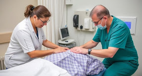
Shoulder Dislocation Rehabilitation Recovery
 |
| Shoulder Dislocation Rehabilitation |
Shoulder Dislocation Rehabilitation
Introduction
A shoulder
dislocation (formally glenohumeral dislocation) is when the
humerus separates from the glenoid cavity of the scapula at the glenohumeral
joint. The shoulder is an inherently unstable joint because it has a
shallow glenoid cavity that only connects with a small portion of the humeral
head.
This
type of dislocation accounts for 50% of primary joint dislocations
and is the most common dislocated joint in the body.
A shoulder
dislocation can be complete or partial, forward (95% of shoulder dislocations),
backward, or downward. The fibrous connective tissue is often stretched or
torn, making it difficult to repair the fracture site.
Causes of Dislocation
The shoulder
dislocates more often than any other joint in the body. Dislocations can
be aggravated by loose or torn fibrous tissues that connect bones. These bones
can be removed with as little force as a gentle tap on the shoulder.
Forceful rotation may cause the humeral head to protrude through the labrum.
Contact sports injuries are common causes of shoulder dislocations,
including those resulting from sports and falls. Trauma from car accidents and
falls can also cause shoulder dislocations.
Human epidemiology
Shoulder dislocation
is the most common large joint dislocation, and dislocation
patterns include anterior dislocation, posterior dislocation,
inferior dislocation, or anterior displacement. Shoulder dislocation
is divided into anterior dislocation and posterior dislocation,
of which anterior dislocation is the most common, accounting for 95% of shoulder
dislocations.
Risk factors for Redis location:
Early dislocation
with poor tissue healing or soft tissue laxity
Young
patients are more active and therefore have a higher incidence of Redis
location...
Patients
with rotator cuff tears or glenoid fractures have a higher incidence of Redis
location...
Mechanism of injury/pathological process
Strong
external force or rapid rotation can cause the humeral head to protrude from
the labrum. Contact sports injuries are a common cause of shoulder dislocations,
as are sports injuries and falls.
Anterior shoulder dislocation
Anterior
dislocations are the most common type of dislocation and occur
when the arm is excessively abducted and externally rotated. In this position,
the subscapular glenohumeral complex acts as an initial traction for anterior
glenohumeral movement. Due to a lack of ligamentous support and dynamic
stability, the glenohumeral joint is prone to dislocation at 90°
abduction and 90° external rotation.
Associated complications and injuries include
Shoulder
instability due to subscapular glenohumeral ligament injury
Hill-Sachs
defect
Bankart
injury or other shoulder labrum injury
Axillary
artery or brachial plexus injury
Posterior
shoulder dislocation (PSD)
Posterior
dislocation is a rare condition that accounts for 3% of shoulder dislocations.
Normally, the humeral head is pushed posteriorly by internal rotation during
the abduction of the arm. Causes include epileptic seizures (most common in
adults, usually bilateral), electrical shock, and trauma during the dislocation
process.
Clinical findings
Anterior
dislocation (the humeral head lies anteriorly, medially, and slightly
below the normal socket and glenoid).
After
acute glenohumeral anterior dislocation:
Reach
out for removal and go to the ER (emergency surgery).
The
normal deltoid contour is lost, and the acromion bulges posteriorly and
laterally...
The
humeral head is palpable anteriorly.
All
movements are restricted and painful.
This
thickness is palpable from below the coracoid toward the axilla.
Injury
to the rotator cuff muscles and bones is possible.
Compression
of the axillary vessels may cause muscle injury, resulting in decreased pulse
pressure and a temporary cold hand.
Peripheral
nerve injuries typically result from traction on the brachial plexus.
Posterior dislocation
In acute posterior glenohumeral dislocation:
The arm
may also be inverted
The
deltoid contour may or may not be lost
The head
of the posterior eminence of the humerus is visible.
Subscapularis
rupture (weakness or inability to internally rotate).
Neurovascular
disorders are rare, but injuries to the labrum and capsule can lead to
posterior shoulder instability.
Late dislocations
are difficult to reduce, so manual reduction should be attempted in
consultation with the treating orthopedic surgeon. Manual reduction is
contraindicated if the shoulder has been dislocated for more than 3
weeks (common in frail elderly patients) or if there is a reverse Hill-Sachs
defect of more than 20% of the joint surface.
Reduction
of posterior dislocations is difficult and should only be attempted in
consultation with the treating orthopedic surgeon. Shoulder dislocation
lasts for more than 3 weeks (common in elderly patients with severe shoulder
dislocation) or more than 20% of the joint surface.
Diagnostic procedure
X-rays
are usually enough to diagnose a shoulder dislocation. However,
computed tomography and MRI are often required to evaluate for glenoid rim
microfractures and ligament/tendon injury.
These
things will come out.
Disabilities
of the Arm, Shoulder, and Hand (DASH).
A quick
DASH
Shoulder Pain
and Disability Index (SPADI).
Numerical
Pain Rating Scale (NPRS).
Management/Interventions
The
dislocated shoulder should be lowered immediately. It is usually
performed in the emergency department after numbness and appropriate pain
medication are administered. There are various methods to lower the shoulder.
See also exercise therapy and shoulder joint therapy.
First dislocation
Treatment
for ASD is usually to reduce the limb and immobilize it for some time (e.g. 6
weeks) to allow the joint capsule to heal properly. To ensure optimal recovery
and eventual return to normal function, systematic physical therapy is required
to reduce muscle atrophy and maintain mobility. During immobilization,
isometric exercises for the shoulder muscles are of great importance.
Surgical repair may be necessary to treat dislocation problems and
related injuries (see above).
For
painful ASDs, the postoperative closure period and the timing of the initiation
of each exercise therapy vary greatly from patient to patient. There is a
paucity of studies comparing the effectiveness of different Rehabilitation
programs, and there is also a lack of evidence to guide postoperative Rehabilitation.
Recent advances in surgical techniques and the diversity of patients presenting
with ASD contribute to these changes [10]. Wang and others proposed a
three-stage protocol.
Stage 1:
Immobilization (up to 6 weeks). Our goal is to work back and forth on
sustainability issues.
Traditionally,
immobilization has been thought to occur in internal rotation, but according to
Miller, external rotation immobilization is more effective because it increases
the contact force between the labrum and the glenoid fossa.
Studies
show that 10° external rotation immobilization has a lower recurrence rate than
10° internal rotation immobilization.
There is
currently no agreement on the duration of immobilization in the sling.
Generally,
individuals under 40 years of age wear the sling for 3 to 6 weeks, while those
over 40 years of age wear it for 1 to 2 weeks of age.
During
the immobilization period, the focus is on the active range of movement (AROM)
of the elbow, wrist, and hand and pain relief. Isometric exercises can be done
with the rotator cuff and biceps. Example: Codman exercise: External rotation
(0-30°) and anterior elevation (0-90°) range of motion (AAROM).
Phase 2
(6-12 weeks): The goal is to regain full range of motion, especially with
external rotation.
Passively
stretch the posterior capsule using joint mobilization and self-stretching
until the full range of motion is restored.
Strengthening
and repetition exercises should not be initiated until the full range of motion
is restored.
Phase 3 (weeks 12-24): Return to sports or daily exercise
Reinforcement
exercises begin. Strengthening exercises should be tailored to the defect.
Strengthening
exercises are usually preceded by mild stabilizing exercises.
Progressive
training should initially focus on the scapular stabilizers, such as the
trapezius, serratus, elevator scapulae, and rhomboids, as well as the rotator
cuff muscles. Then we move on to the larger muscle groups like the deltoids,
latissimus dorsi, and pectoralis major.
We have
begun to emphasize functional exercises, including proprioceptive training, to
increase patient activity and social participation.
Related topics: Back to the game within the game
Differential diagnosis
Fractures
(clavicle, glenoid, humeral head, tuberosity, proximal humerus)
Rheumatoid
arthritis
Rotor
cuff injury
Acromioclavicular
joint dislocation
Labral
lesion
Shoulder
subluxation
Axillary/suprascapular
nerve palsy

